Thermal imaging makes it easier to solve many problems of doctors and specialists in health improvement: it allows to predict the course of the disease, to determine the nature of the process and the degree of its prevalence, damage to certain organ structures.
This harmless and comfortable method has proven its effectiveness in early diagnosis and prevention of mastopathy and breast cancer. With the help of VedaPulse thermal imaging diagnostics (Thermography software module), a naturopath can carry out a detailed assessment of the breast, detect diseases in their early stages, prevent or monitor the effectiveness of treatment of dyshormonal diseases of the mammary glands.
Thermal imaging diagnostics allows us to assess the change in tissue temperature, which is associated with biochemical features that precede structural changes in tissues. Biochemical, immunological and enzyme immunoassay diagnostic methods allow a doctor or a naturopath to assume the presence of a malignant process. An important advantage of modern thermal imaging is the visual "thermal navigation" of the location and volume of the process and the measurement of the dynamics of temperature changes in "cold" or "pharmaceutical" samples, as well as the computer overlay - "morphing" - of images with photos and structural studies.
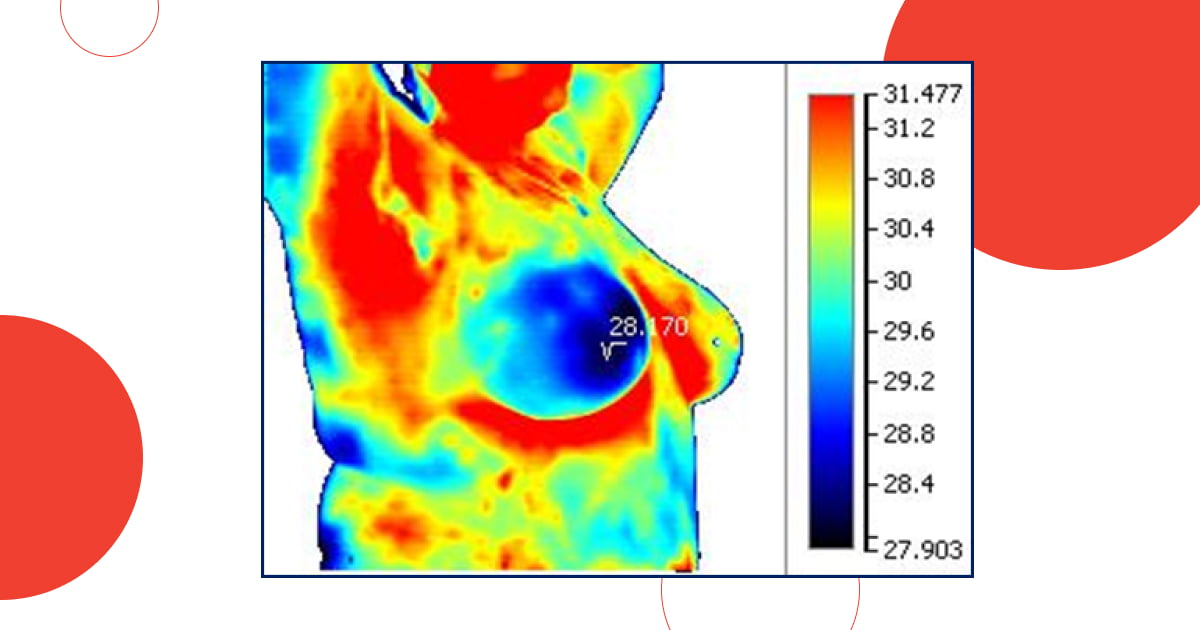
Let's look at clinical examples of the use of medical thermography in the diagnosis and monitoring of mastopathy. The main diagnostic criteria are the asymmetry of the temperature field of the mammary glands, the presence of foci of hypo- and hyperthermia.
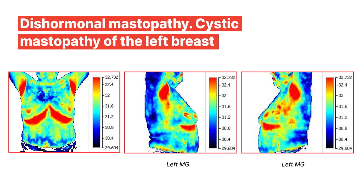
1. Dishormonal mastopathy. Cystic mastopathy of the left breast
Mammary glands are symmetrical. Inhomogeneous hypothermia of the parasternal regions, in the upper-medial quadrants of the mammary glands.
Axillary areas are thermosymmetric. Severe hyperthermia of the periareolar areas. In the right gland there are hyperthermic areas along the ducts of the gland. On the left side there are multiple foci of inhomogeneous hyperthermia along the course of the regional lymph nodes. In the upper lateral quadrant on the left there are two distinct foci of hyperthermia with clear borders. Eosinophils predominate in one of the foci of hyperthermia on the left, which is more typical of a cyst.
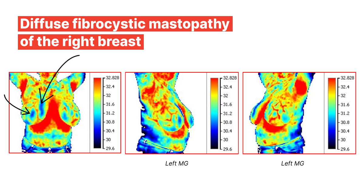
2. Diffuse fibrocystic mastopathy of the right breast
The right mammary gland is slightly larger than the left one.
In the right lateral projection, there is a lacuna-like hyperthermia of the interlobular lymph vessels with an inhomogeneous hyperthermia course into the axillary group of lymph nodes.
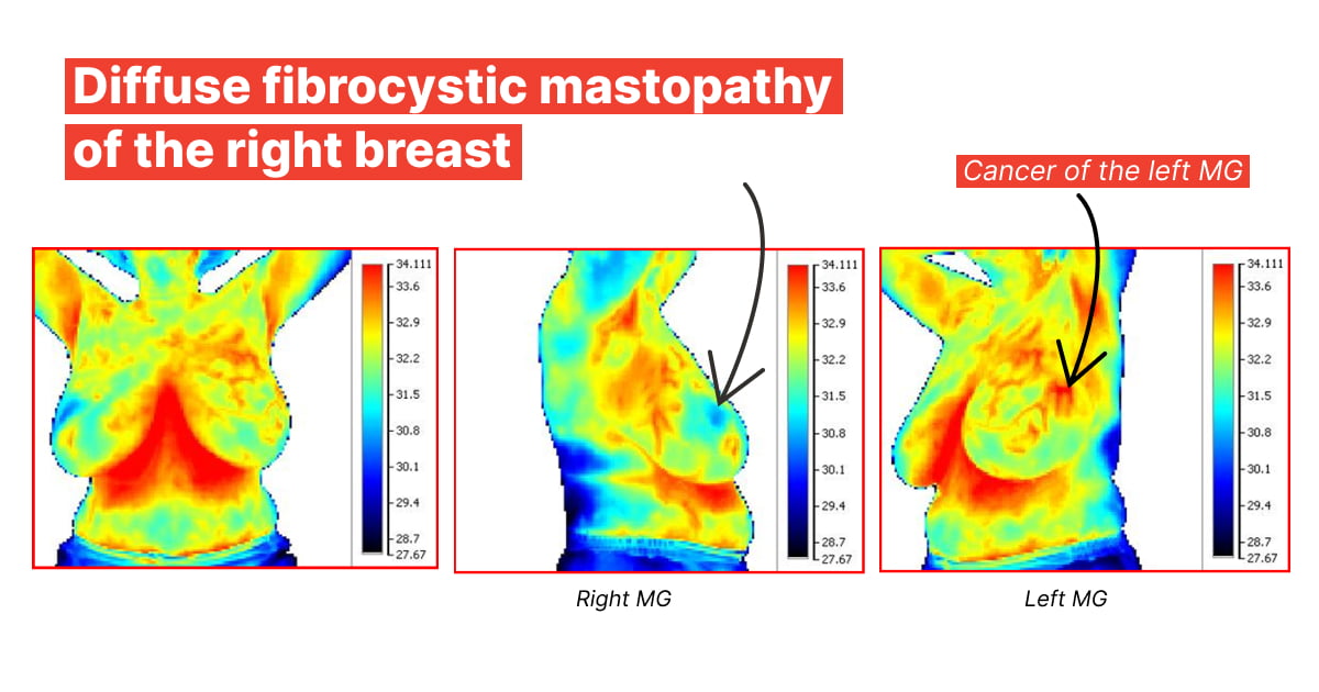
3. Diffuse fibrocystic mastopathy of the right breast
The glands are slightly asymmetrical (the left one is pulled up when the arms are raised).
On the left there is a pronounced enhancement of the contour of the lymphatic ducts in the direction of the parasternal lymph nodes.
Foci of hyperthermia in the projection of the supra- and subclavian lymph nodes on the left, in the upper lateral quadrant.
On the left there is a zone of homogeneous hyperthermia in the form of a capital Greek letter "eta", which does not coincide in length with the course of the vessels and lobules of the gland. The perifocal hyperthermia extends into the axilla.
Clinical examples show how diagnostics with thermal imaging and Thermography software module can help doctors and healthcare specialists in their practice. Using Thermography, you will be able to make a more accurate and reliable assessment, determine risks and prescribe prevention of mastopathy and other diseases of the mammary glands. Remember that the patient needs to see a doctor to confirm or exclude the diagnosis.
Thermal imaging control is a reliable way to detect injuries in time, take measures and preserve health. The method has proven its effectiveness not only in functional diagnostics of various inflammatory processes of internal organs and tissues, but also as a means of monitoring patient dynamics in sports medicine, physiotherapy and cosmetology.
Contact VedaPulse managers so as to learn more about the method and its application, get clinical examples and data from recent studies on the effectiveness of thermal imaging diagnostics.